Podcast
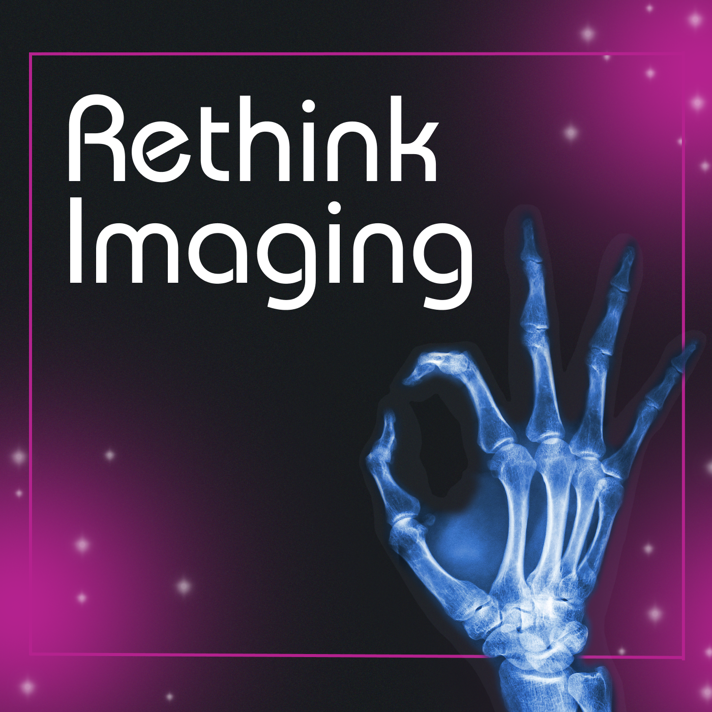
Rethink Imaging
Hosted by Imalogix · EN
Rethink Imaging is a podcast that delves into the fascinating world of radiology, offering a fresh perspective on medical imaging and its impact on patient care and healthcare outcomes. The podcast, hosted by Chris St John, aims to bridge the gap between complex radiological concepts and the curious mind, from seasoned professionals to radiology enthusiasts.
Each episode features engaging discussions with leading experts in medical imaging, including radiologists, medical physicists, radiologic technologists, world-leading academics, and industry thought leaders. Through inspiring interviews, the podcast explores a wide range of topics, from advancements in imaging technology and diagnostic practices to the future of radiology in healthcare.
30episodes
Episodes
Newest firstAll episodes
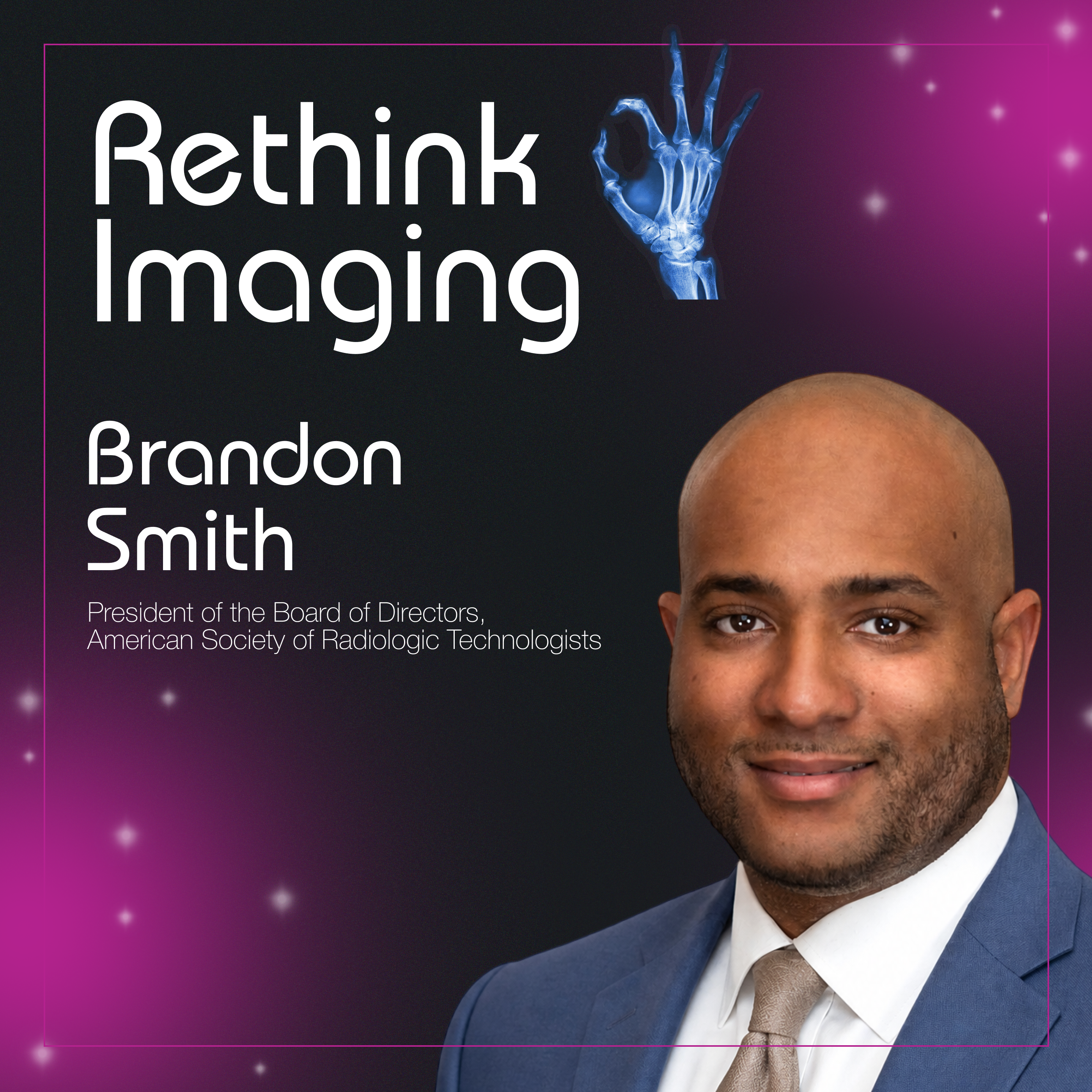
Medical Imaging Professionals Need More Than Recognition: They Need Influence with Brandon Smith
4d ago00:47:09Tap to summarizeMedical imaging professionals and radiation therapists generate massive hospital revenue and deliver critical direct patient care, yet they are frequently classified as "technical personnel" or "other" staff. How does a vital healthcare sector shift from being quietly dissatisfied to completely unignorable?In this episode of Rethink Imaging, host Chris St. John is joined by Brandon Smith, a cardiovascular/IR technologist, holder of an MS in Radiological Sciences and an MBA, and the first person in history to serve consecutive terms as president of the ASRT. Brandon breaks down the profound difference between simply occupying a committee seat (volunteerism) and executing real operational impact (advocacy). He shares the tactical playbook behind the ASRT's massive "Be Seen" national ad campaign during the Olympics, details how the profession mobilized 42,000 signatures in just one weekend to petition the OMB for professional reclassification, and explains why workforce shortages and scope encroachment are not static problems to be solved, but ongoing environmental factors that require constant, measurable clinical advancement.What You’ll Learn:The Advocacy vs. Volunteerism Split: Why showing up and filling a seat on a committee is just the baseline, and how true advocacy must result in tangible policy and classification changes.The Theory of Mobilization: A functional five-step framework (Visibility, Recognition, Appreciation, Advocacy, Advancement) to systematically elevate a profession's cultural and operational value.Reclassifying the RT: The operational and legislative reality behind pushing the Office of Management and Budget (OMB) to move radiologic technologists from a "hyper-technical" status to recognized "professionals."The Shortage & Encroachment Factors: Why cyclical staffing shortages and cross-lane scope encroachment are predictable environmental factors rather than standalone crises, and how to build structural resilience against them.Shifting from Consumption to Production: Why medical imaging must aggressively produce its own peer-reviewed literature and citations rather than borrowing clinical authority from external medical personas.Chapters:[00:02:18] The Familial Route: Tracing a path from an engineering detour and a neuroimaging externship back into the family legacy of medical imaging.[00:05:51] A Shift in Perspective: How the perception of radiologic technology evolved from a laborious contingency job into a rich, intentional career.[00:07:25] The Trap of the Contingency Plan: Confronting the harmful narrative of imaging as an "ancillary" backup plan and refusing to accept residence in dissatisfaction.[00:09:47] Complacency vs. Worthiness: Analyzing why revenue-generating service lines allow themselves to be drowned out by commercially-facing hospital personas.[00:14:23] Policy Over Banners: Why real professional appreciation must show up in concrete policy, procedures, and minority seats at executive decision-making tables.[00:18:02] Overcoming Volunteerism: The critical distinction between individual pageantry/plaques and collective professional impact.[00:21:36] Forest vs. Tree (Modality Mentality): Why specialists must celebrate their specific modality while maintaining fierce purpose for the broader medical imaging profession.[00:26:58] Mobilizing 42,000 in a Weekend: Inside the high-stakes push to petition the OMB for professional reclassification before the 10-year window closed.[00:30:00] The "Be Seen" Campaign: How a board discussion transformed into a national Olympic ad campaign reaching 27 million people.[00:37:11] The Cyclical Shortage Factor: Moving away from reactive panic over CT and radiography vacancies to focus on long-term infrastructure.[00:40:34] Producing vs. Consuming Data: Addressing the research deficit and the urgent need for RTs to publish peer-reviewed, citable literature.[00:42:40] The Unprecedented Second Term: The behind-the-scenes mechanics of an emergency special election and choosing continuous team momentum over personal pageantry.Rethink Imaging Podcast is handcrafted by our friends over at: fame.so
Transcribe →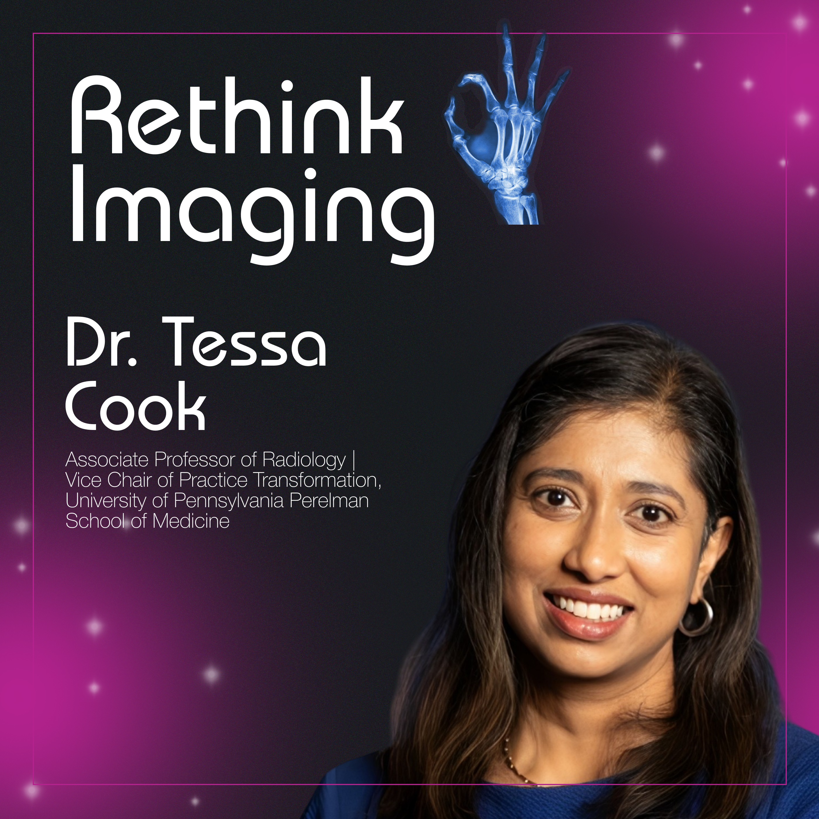
Why Most Radiology AI Fails: Workflow, Governance & Integration Problem with Tessa Cook
3w ago00:42:20Tap to summarizeAdvanced engineering and deep learning can create breathtaking diagnostic models, but how do we successfully integrate complex AI tools into a high-volume, multi-site hospital system without creating more chaos?In this episode of Frame by Frame: Rethink Imaging, host Chris St. John sits down with Dr. Tessa Cook, a cardiovascular radiologist, national leader in imaging informatics, and Commission Chair for Informatics for the American College of Radiology (ACR), to break down the reality of clinical technology adoption. Dr. Cook explains the profound structural difference between a model that performs well in a retrospective testing lab and one that actually adds value to a radiologist’s busy shift.She shares concrete case studies detailing how her team built "Arnie" (Automated Radiology Recommendation Tracking Engine) to bridge fragmented patient care, ultimately giving rise to Penn Medicine's enterprise-wide High-Risk Follow-Through program that caught 12 early-stage cancers in its initial phase. We also look at the industry-wide hurdles to scaling medical AI, analyzing Penn's comprehensive three-phase evaluation framework, the evolution of clinical governance, and why standard technical metrics fail when algorithms completely lack access to crucial patient clinical context.What You’ll Learn:The Workflow Commandment: Why the absolute highest-performing AI algorithm is completely useless if it cannot seamlessly connect to a radiology practice's existing daily workflow.The Three-Phase AI Evaluation: How Penn Medicine rigorously tests new models—moving from retrospective performance metrics to limited prospective user experience testing before making a purchase.The Clinical Context Gap: Why pixel-based AI tools experience high discordance rates when forced to evaluate medical imaging in complete isolation from a patient’s full medical history and lab results.Closing the Follow-Up Loop: How mining structured data elements allows automated tracking engines to nudge clinicians and prevent critical downstream diagnoses from falling through the cracks.Reimaging ROI in Healthcare: Why the return on investment for clinical technology must look beyond simple dollars to measure cognitive burden reduction, clinician burnout, and direct lives saved..Chapters:[00:00] The Core Informatics Problem: Why workflow integration dictates the success or failure of even the best clinical AI.[02:00] Leadership Lessons from the Kitchen: An unexpected look at how Gordon Ramsay’s high-pressure team dynamics mirror healthcare operations.[05:43] The Engineer-Physician Trajectory: Combining computer science, bioengineering, and medicine to tackle complex workflow obstacles.[07:55] Defining Practice Transformation: Shining a light on systemic healthcare problems to optimize patient and clinician experiences.[08:47] Going Back to 2010: Developing the open-source Radiance dose tracking software during residency to combat radiation overexposure.[13:30] The Flaws of Pennsylvania Act 112: Analyzing the real-world operational challenges of mandated patient test notifications.[16:38] Fragmented Care & Information Exchange: Why a lack of unified data sharing between healthcare systems allows critical follow-ups to get missed.[18:10] Engineering "Arnie": Building an automated recommendation tracking engine using discrete data elements long before the LLM boom.[20:10] 12 Lives Saved: Measuring the profound clinical success of Penn Medicine's enterprise-wide High-Risk Follow-Through program.[21:23] Redefining the ROI of Healthcare AI: Looking past financial metrics to evaluate safety, efficiency, and cognitive burden reduction.[23:58] Penn's 7-Year AI Governance Evolution: Streamlining rigorous validation models and achieving the ACR's ArchAI center designation.[26:51] A Three-Phase Evaluation Strategy: Breaking down retrospective verification, limited prospective testing, and user experience deployment.[28:53] Discordance vs. Ground Truth: Navigating the nuances of imaging measurements and the vital role of patient outcomes.[30:11] The Missing Data Points: Why pixel-only AI algorithms fail to interpret imaging with the nuance of a specialized radiologist.[32:52] Training the Next Generation: Graduating over 100 alumni from Penn’s elite Imaging Informatics Fellowship.[37:16] An Apologetic Shift Toward AI: Balancing modern generative AI tools with foundational imaging standards like DICOM and HL7.[39:23] The Value of Society Governance: Key takeaways from serving as Chair of the Society for Imaging Informatics in Medicine (SIIM).Rethink Imaging Podcast is handcrafted by our friends over at: fame.so
Transcribe →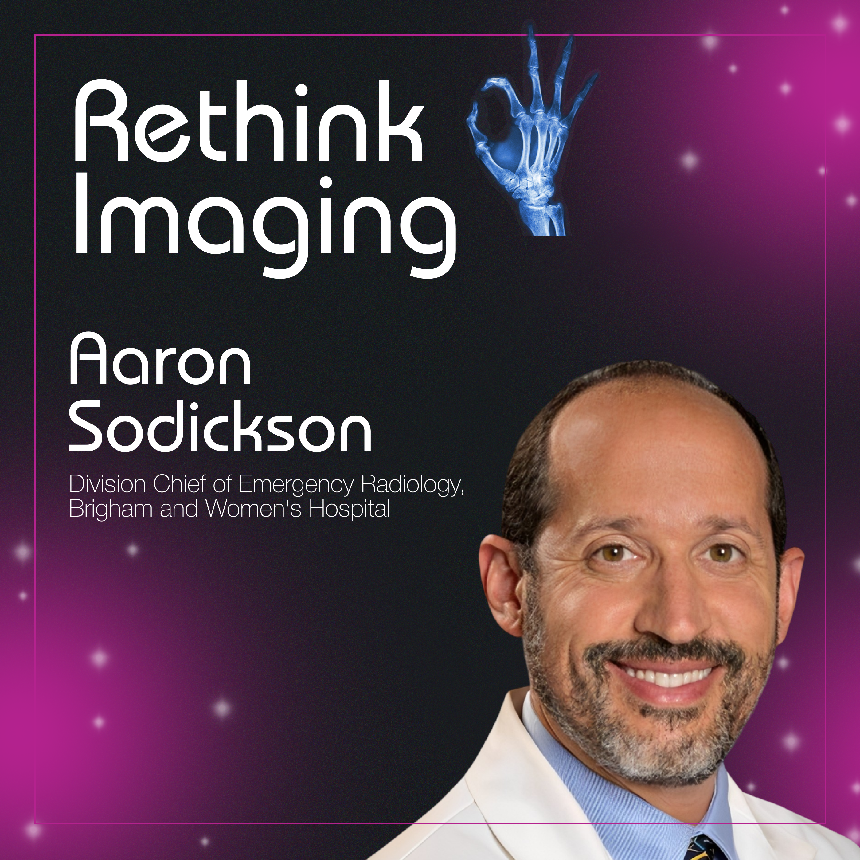
Photon Counting CT Could Revolutionize Diagnostic Radiology
Jun 2500:40:39Tap to summarizeAdvanced engineering can create breathtaking diagnostics, but how do we successfully bring complex imaging technology out of a development lab and make it work in a high-volume emergency room?In this episode of Rethink Imaging, host Chris St. John sits down with Dr. Aaron Sodickson, Division Chief of Emergency Radiology at Mass General Brigham and director of CACTI, to break down the mechanics of photon counting CT. Dr. Sodickson explains the profound structural difference between older energy-integrating detectors and modern semiconductor arrays that record individual electrical pulses.He shares concrete case studies detailing how this technology drops patient radiation doses by 30% to 50%, yields beautiful point-two-millimeter resolution for subtle bone fractures, and completely automates color-coded iodine mapping to clear incidental findings on the spot. We also look at the industry-wide hurdles to scaling this tech, including the fight for unified DICOM standards inside standard hospital PACS software.What You’ll Learn:The Scintillator Leap: How skipping the step of converting X-rays into light removes pixel septa and maximizes dose efficiency.The 50% Noise Reduction: Why setting energy thresholds allows scanners to toss out electronic noise and protect patients.Definitive ER Troubleshooting: Differentiating benign calcifications from active brain hemorrhages during the initial patient scan.The Last Mile Problem: Working alongside the AAPM and PACS vendors to embed real-time quantitative measurement tools into everyday hospital software.Chapters:[00:00] The Emergency Room Mandate: Managing intense time pressures across a multi-site trauma network.[03:53] Kicking the Tires at CACTI: Overcoming the operational hurdles of the "last mile" translation gap.[08:08] Conventional vs. Dual-Energy CT: Utilizing multiple energy spectra to isolate material properties.[09:20] The Photography Automatic Trap: Why multi-million dollar scanners are underutilized across the country.[11:50] Four Game-Changing Wins: Isolating pathology, strengthening protocols, dropping dose, and ending follow-up loops.[12:54] Differentiating Calcium vs. Hemorrhage: Clearing emergency headache and trauma cases upfront.[16:36] Automating the Background Process: Creating background calculations to deliver automated colored data to PACS.[20:42] Borrowing Technology from CERN: The direct integration of particle physics into clinical imaging chains.[29:12] The Beauty of Ultra-High Resolution: Achieving 0.2-millimeter slice details to view subtle micro-fractures.[31:36] Fixing Pulmonary Embolus Protocols: Using low-energy processing to eliminate scanner timing failure rates.[33:02] The Fight for New DICOM Standards: Forcing an alliance between the AAPM, manufacturers, and software vendors.[37:37] Advice for the Next Generation: Why radiology trainees must sit with technologists to master the real console knobs. Rethink Imaging Podcast is handcrafted by our friends over at: fame.so
Transcribe →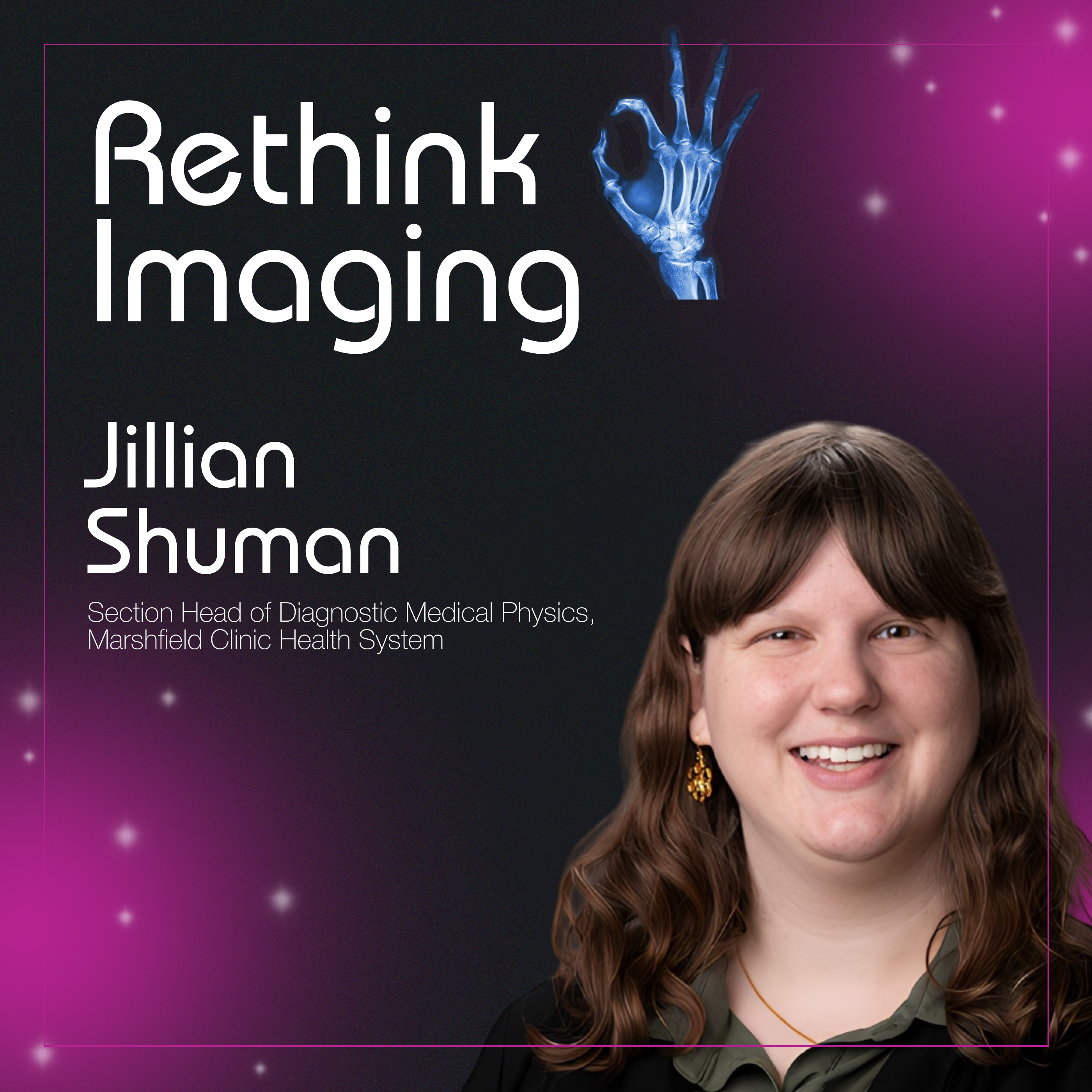
Medical Physics in Rural Hospitals: Higher Dose, Lower Resources, Bigger Stakes
Jun 1100:36:52Tap to summarizeThe world of medical imaging is often discussed through the lens of cutting-edge academic research, but the majority of America's healthcare landscape relies on rural facilities operating on razor-thin margins. Jill Shuman, DMP, joins the podcast to share her boots-on-the-ground experience driving through a multi-hour radius to keep local scanners compliant, safe, and operational.Jill shares the harsh reality of spending years on "break-fix" budgets, where critical imaging equipment can only be replaced if it is entirely beyond repair—a cycle that leads to higher radiation doses for patients and increased system downtime. The conversation dives deep into the administrative and logistical hurdles unique to rural settings, including the influx of traveller technologists who frequently miss scanning fundamentals, the lack of rideshare infrastructure preventing patient access, and a punishing reimbursement system that creates a financial catch-22 for struggling community hospitals. It’s a powerful look at how local healthcare advocates fight for their neighbors, proving that personalized medicine in rural America is built on deep communal bonds.What You’ll Learn:The Break-Fix Cycle: How restrictive equipment budgets unintentionally lead to higher CT doses and increased scanner downtime.The Traveler Tech Paradox: The clinical and image quality risks that emerge when rural hospitals are forced to staff facilities 100% with temporary agency technologists.The Rural Catch-22: How CMS dose metrics and reimbursement penalties disproportionately punish underfunded hospitals with older equipment.Innovating on a Budget: How the Marshfield Clinic became the first in Wisconsin to adopt contrast-enhanced mammography, drastically lowering patient costs while matching MRI accuracy.The Reality of Access: Why the closure of rural hospitals creates devastating gaps in community health, and how physicists ground themselves in the human story behind the data..Chapters:[00:00] The Precariousness of Rural Healthcare: Jill shares the sobering reality of driving past a newly shuttered local hospital on her daily commute, highlighting the fragility of small-town medicine.[03:34] The Three Hats of a Rural Physicist: From splitting time between equipment testing and compliance paperwork to the daily "detective work" required to troubleshoot unexpected clinical issues.[06:52] Medical Physics 3.0 in Small Towns: Overcoming the industry misconception that outlying facilities cannot provide deeply personalized, high-level clinical care.[10:49] Budget Barriers and the Traveler Influx: How thin financial margins force rural clinics to balance lower patient volumes against the training risks of short-term agency staff.[13:30] The Break-Fix Reality: Jill details the hidden costs of aging machines and shares a case study of immediate image quality issues caused by a 100% traveler workforce.[16:12] No Uber, No Lyft: Navigating severe transportation hurdles and geography to get patients to specialized imaging appointments.[18:01] Entering the "Donut of Truth": Discussing high-utilizer CT scans, emergency room dynamics, and the rise of medical imaging meme culture on social media.[23:45] The Reimbursement Trap: Why penalizing higher-dose, older scanners via federal metrics hits thin-margin community centers the hardest.[27:44] Celebrating Huge Wins: How the Marshfield Clinic became the first hospital in Wisconsin to adopt low-cost, high-accuracy contrast-enhanced mammography.[33:40] Shouting for Resources: A final plea for financial grants, structural support, and updated protocol tools from the AAPM to assist rural physicists.Rethink Imaging Podcast is handcrafted by our friends over at: fame.so
Transcribe →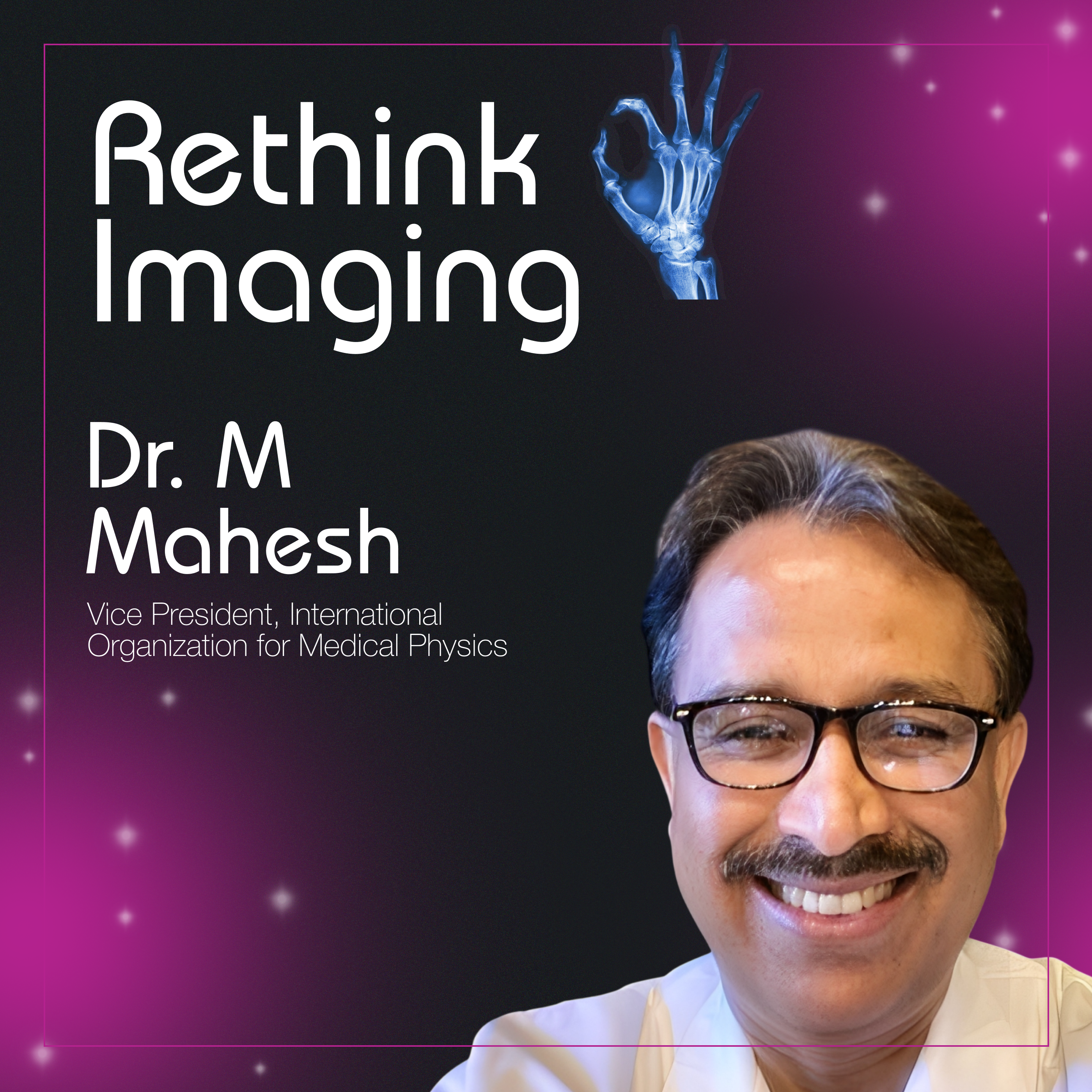
Are CT Scans Safe? What Most People Get Wrong About Radiation
May 2800:53:49Tap to summarize The fear of radiation is often rooted in what we cannot see, smell, or touch, leading the public to associate life-saving CT scans with historical disasters like Hiroshima or Chernobyl. Dr. M. Mahesh, a Professor of Radiology and clinical leader, joins the podcast to combat this "infodemic" of medical disinformation.Dr. Mahesh shares his experience leading national reports on radiation exposure, explaining why a reported "600% increase" in the US population's dose was a misunderstood statistic that triggered unnecessary Congressional hearings. He dives into the counterintuitive science of why patient shielding, once a standard practice, can actually increase radiation doses due to modern automatic exposure controls. Beyond the physics, the episode explores the "long game" of building public trust through social media, the role of technologists as the "window for radiology," and how experts can "flood the zone" with good information to ensure AI models provide accurate medical adviceWhat You’ll Learn:The 600% Myth: How statistics about US radiation exposure were sensationalized and the reality of the 20% decrease seen in more recent years.The "Light Bulb" Analogy: Why an X-ray room holds zero radiation the second the machine is turned off.The Shielding Paradox: Why lead aprons can sometimes obscure clinical images or force machines to output more radiation.Combatting "Clickbait" Science: Strategies for clinicians to use social media and "comic book" storytelling to simplify complex safety concepts.The Role of AI: Why professionals must engage online to ensure Large Language Models (LLMs) prioritize accurate medical data over "scare tactics".Chapters:[00:00] The Headline That Horrified Congress: Deconstructing the 600% radiation increase report.[01:55] The Professional Obligation: Why Dr. Mahesh steps out of the academic "ivory tower" to speak to the public.[06:38] Communicating in "Multiple Languages": Explaining radiation to everyone from radiologists to grandmothers.[11:53] The MAMO Scare: How local news cycles turn life-saving screenings into fear-based clickbait.[23:26] Radiation Myths: Addressing the "Hiroshima reference point" and the light bulb analogy.[31:22] The Window of Radiology: Why technologists are the most important communicators in the clinic.[37:01] The Death of Shielding: The scientific evidence against using lead aprons for patients.[48:12] Advice for Professionals: How to start your own journey in public advocacy. Rethink Imaging Podcast is handcrafted by our friends over at: fame.so
Transcribe →
Killing Tumors Without Chemo: Sci-Fi Treatments Already in Hospitals with Dr. Mina Makary
May 1400:30:24Tap to summarizeThe gap between medical possibility and patient awareness has never been wider. In this episode, Dr. Mina Makary, a leader in clinical informatics and interventional radiology at The Ohio State University, dismantles the idea that cancer treatment always requires systemic chemotherapy or invasive surgery.Dr. Makary introduces Histotripsy, a groundbreaking technology that uses ultrasound waves to mechanically destroy tumors without even a needle poke. The conversation expands beyond oncology to address everyday quality-of-life issues: treating knee arthritis with Genicular Artery Embolization to avoid joint replacement, and providing non-surgical solutions for uterine fibroids and prostate enlargement. This episode is a tactical roadmap for patients and providers looking to leverage technology that treats disease at its "doorstep" while preserving the body’s integrity.What You’ll Learn:Interventional Oncology (IO): Why IO is now considered the "fourth pillar" of cancer care alongside medical, surgical, and radiation oncology.Histotripsy Explained: How ultrasound-induced "cavitation" can kill a tumor non-invasively, leaving no mark behind.The "Doorstep" Delivery: The mechanics of Y-90 and TACE, delivering radiation or chemo directly to a tumor while sparing the rest of the body.Chronic Pain Solutions: How blocking abnormal blood vessels (GAE) can provide up to 24 months of relief for knee arthritis.Uterine and Prostate Health: Why "minimally invasive" shouldn't just be an option, it should be the first trial before major surgery.Chapters:[00:00] The Future is Now: Introduction to Histotripsy and needle-free tumor destruction.[03:15] The Fourth Pillar: Defining Interventional Oncology and the "doorstep" treatment model.[05:35] The Band-Aid Cure: The mechanics of freezing or heating tumors in real-time.[08:59] Cavitation vs. Heat: The technical benefits of ultrasound waves over traditional ablation.[11:34] Moving Beyond Wear and Tear: A new way to treat knee pain by targeting inflammation, not just bone.[17:23] Sparing the Uterus: Why uterine fibroid embolization (UFE) is a "natural" surgical alternative.[22:17] Men’s Health & BPH: Shrinking the prostate without the risks of traditional surgery.[27:44] The AI Integration: How chatbots and predictive algorithms are supporting the IR workflow. Rethink Imaging Podcast is handcrafted by our friends over at: fame.so
Transcribe →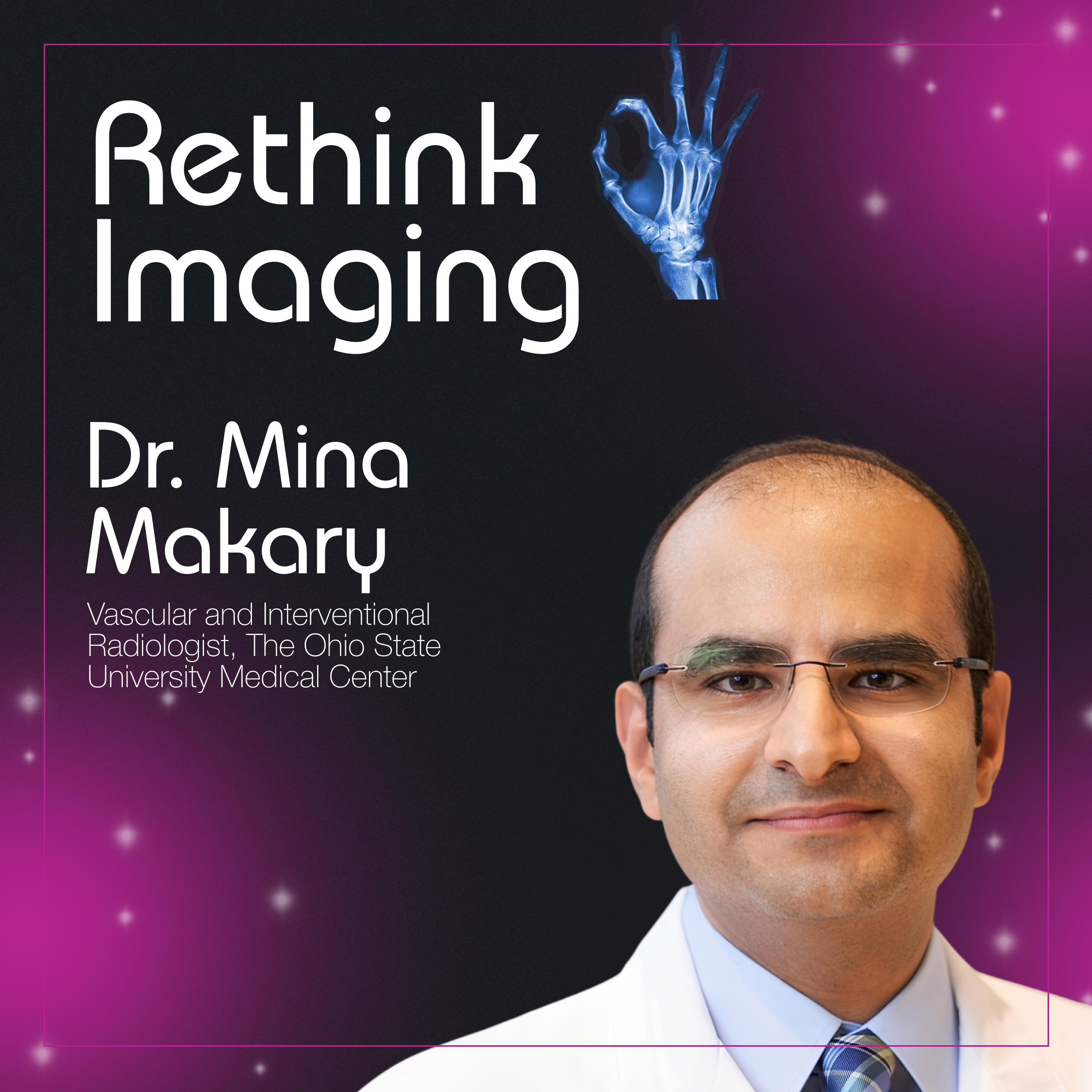
It’s Not Just Burnout: What’s Also Affecting Radiologists with Dr. Mina Makary
Apr 3000:39:29Tap to summarizeThe landscape of medical imaging is shifting from simple diagnosis to active, minimally invasive treatment, yet many patients and primary care providers remain unaware of these life-changing options. Dr. Mina Makary joins Rethink Imaging to bridge this awareness gap, detailing his journey in building a pioneering inpatient IR service at Ohio State.The conversation dives deep into the "art of IR," where complex venous reconstructions and targeted cancer treatments offer hope to patients who have been told they have no other options. However, providing this high-level care comes with a heavy emotional price. Dr. Makary discusses the concept of "moral injury", a state of disenfranchisement that occurs when physicians are forced to navigate bureaucratic and economic conflicts that compromise patient care. This episode serves as a call to action for both institutional change and better public education to ensure that neither the doctor nor the patient is left behind by a system under stress.What You’ll Learn:The Identity of IR: Why Interventional Radiology was recognized as a distinct specialty in 2012 and how it differs from diagnostic imaging.Building a Service from Scratch: The tactical challenges of launching an IR inpatient team, from financial proposals to recruiting the right "mindset".The "Hammer and Nail" Trap: Understanding the importance of multidisciplinary collaboration over "turf wars" in medicine.Moral Injury vs. Burnout: Why systemic "moral injury" leads to permanent damage and why individual wellness acts aren't enough to fix it.Patient Advocacy: How to empower patients to seek minimally invasive treatments like uterine fibroid embolization and tumor ablation.Chapters:[00:00] Defining Moral Injury: The difference between repeated stress and the trauma of being unable to do the "right thing."[03:11] The Awareness Gap: Why the public and even other doctors often don't know what IR can offer.[08:22] Launching a Legacy: The process of building an APP-led inpatient service at a major academic center.[13:08] The IR Toolbox: A look at life-saving procedures, from stopping traumatic bleeds to freezing tumors.[17:13] Complex Venous Work: Giving hope to "disabled" patients through creative problem solving in vascular reconstruction.[25:37] Building Bridges: How to communicate with referring physicians without "undermining" their authority.[32:04] The Wellness Myth: Why you can't "yoga your way" out of a broken healthcare system. Rethink Imaging Podcast is handcrafted by our friends over at: fame.so
Transcribe →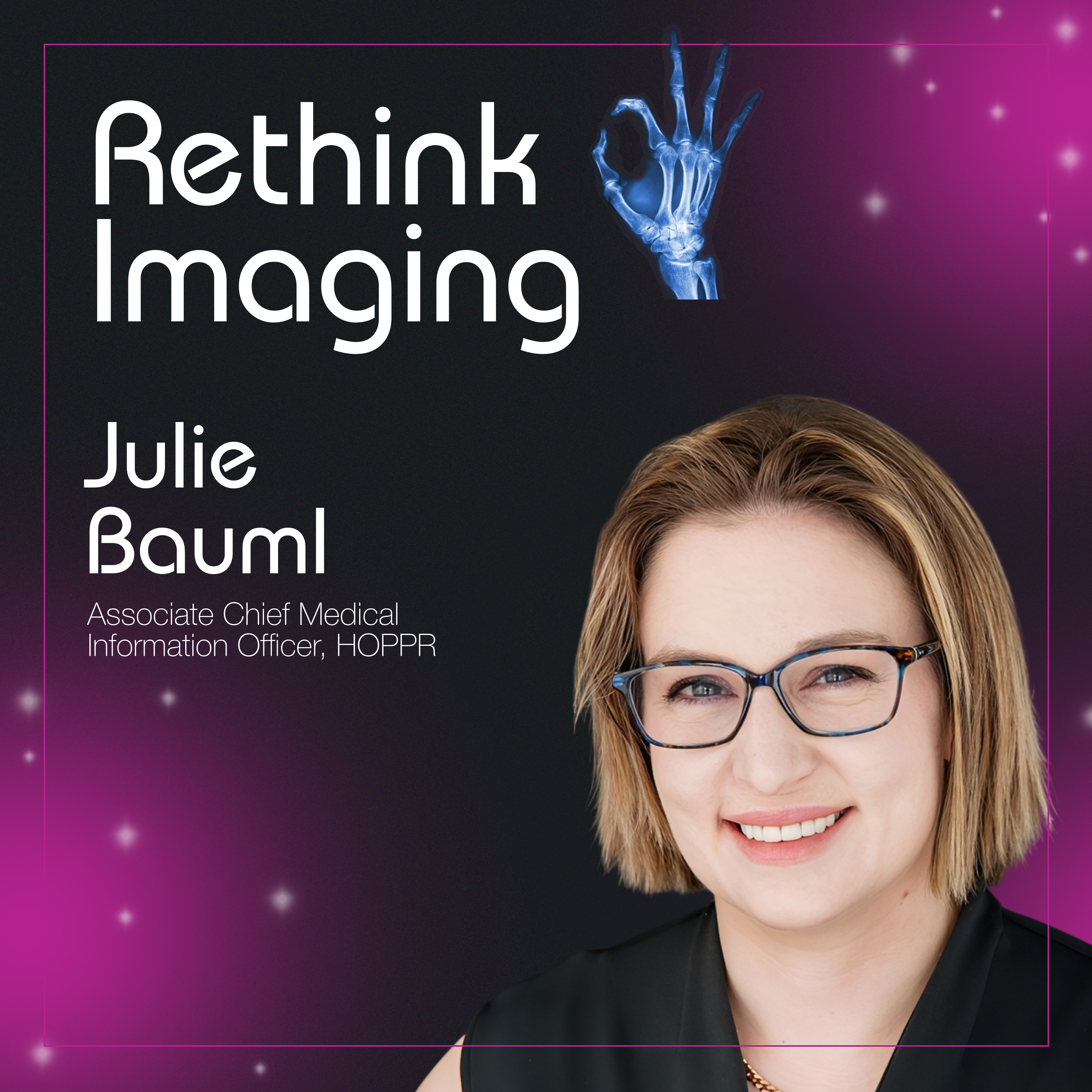
The Cognitive Ceiling: Why AI Must Support Radiologists, Not Replace Them
Apr 1600:45:48Tap to summarizeThe field of radiology is at a breaking point, facing a massive "volume crush" that threatens to push professionals toward early retirement. Dr. Julie Bauml, a board-certified radiologist who transitioned into clinical informatics, joins the show to discuss why technology must be reframed as a tool for augmentation rather than a replacement for human expertise.The episode explores the "cognitive ceiling", a biological limit on how many high-level decisions and visual inputs a brain can process in a day. Dr. Bauml shares sobering anecdotes of AI tools implemented without radiologist input, resulting in poor workflow integration and even decreased reimbursement. From the "Minority Report" dream of hands-free, spatial computing to the nuances of "ground truth" in clinical data, this conversation is a tactical roadmap for how to keep radiologists meaningfully in the loop while leveraging AI to handle the rote, draining tasks that lead to burnout.What You’ll Learn:The Cognitive & Visual Ceiling: Understanding the biological limits of human image interpretation and decision-making.Augmentation over Replacement: Why the 10-year-old narrative of "obsolete radiologists" failed and where the industry is moving now.The "Non-Doctoring" Tasks: Identifying workflow bottlenecks—like chart searching and window alignment—that AI is best suited to solve.The Ground Truth Complexity: Why "weak labels" and human variability make training medical AI harder than standard machine learning.Spatial Computing in Radiology: How VR and AR could solve ergonomic injuries and liberate radiologists from the "giant monitor" setup.Chapters:[02:24] The Career Pivot: Transitioning from clinical practice to informatics to save the "spark" of radiology.[04:05] The Volume Crush: Why the fee-for-service structure and high volume create an interdependent crisis.[06:01] Defining the Ceiling: The visual cortex limit—you can't just "tape your eyes open" to read more.[09:43] The Replacement Myth: Why early AI "point solutions" alienated the workforce.[14:39] Seat at the Table: The importance of including MDs in industry leadership and product design.[19:11] Implementation Failures: An anecdote on how poorly integrated tools can actually make a backlog worse.[25:03] Ground Truth & Data Quality: Why "more data" isn't the answer to baking a better clinical "cake".[41:00] Spatial Computing: The future of hands-free radiology and reading cases from anywhere.Rethink Imaging Podcast is handcrafted by our friends over at: fame.so
Transcribe →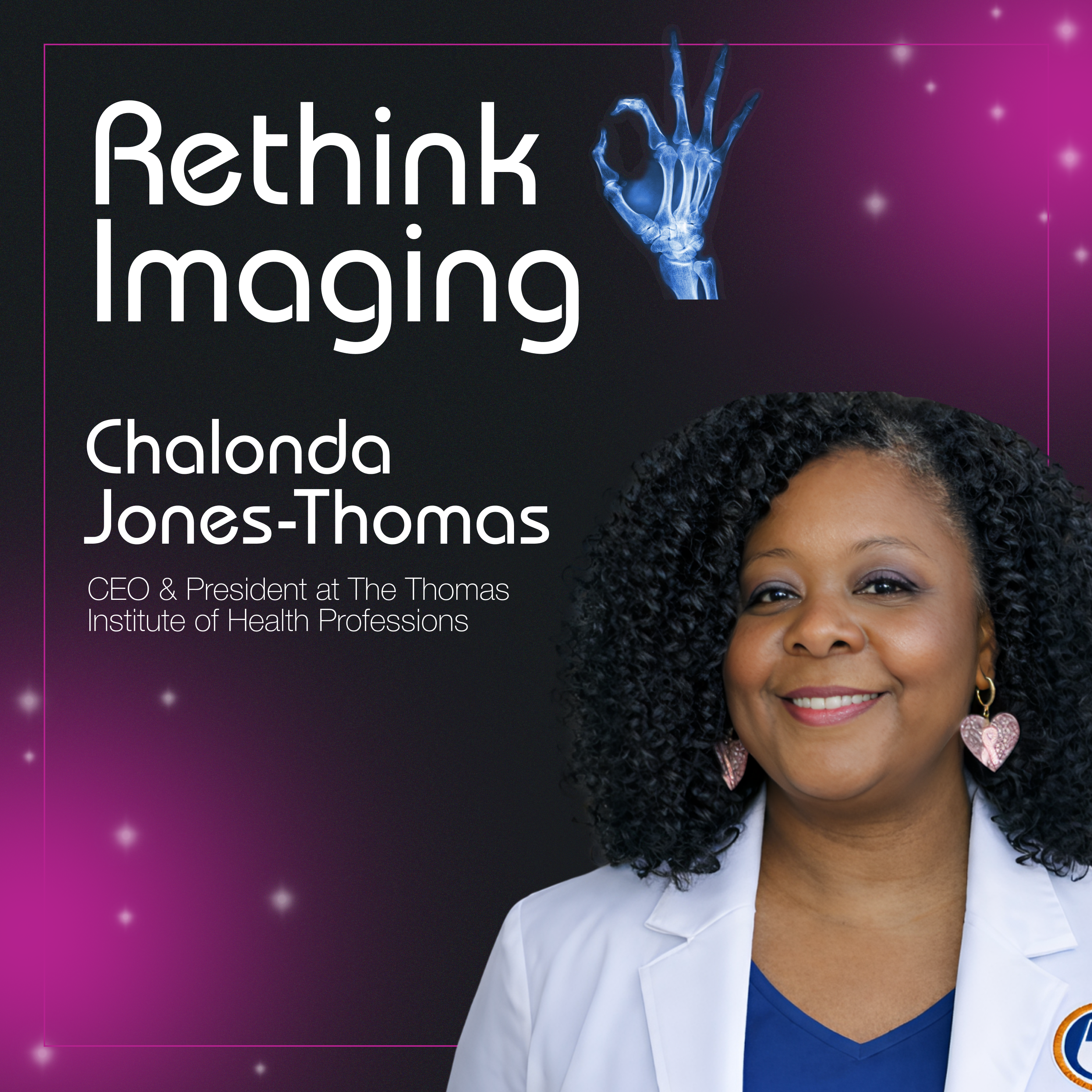
The Triple-Certified Technologist: Fixing Radiology’s 20% Workforce Gap
Apr 200:39:48Tap to summarizeThe medical imaging workforce is facing a critical shortage, with CT tech vacancy rates reaching nearly 20%. Chalonda Jones-Thomas identifies a primary cause for the struggle: a disconnect between outdated textbooks and the rapid advancement of real-world imaging technology. To address this, Chalonda developed the first fully online CT and MRI training programs designed specifically for medical imaging professionals.The episode explores the operational and pedagogical shifts required to move training into a digital space. Chalonda explains how her programs use video, simulation software, and rigorous checkpoints to ensure students are clinically competent before ever touching a patient. By removing geographical barriers, these programs provide a lifeline for technologists in rural areas and military personnel stationed overseas who need civilian-ready credentials. Chalonda reframes online education not as a "hands-off" experience, but as a flexible, data-driven system that empowers technologists to upskill without putting their lives or careers on hold.What You’ll Learn:The Textbook Lag: Why traditional education materials are falling behind the rapid pace of CT and MRI technology.Proving Online Integrity: How simulation software and rigorous assessments won over skeptical radiologists and directors.Empowering the Underserved: How online education provides advanced imaging paths for rural technologists and working parents.Military-to-Civilian Transition: The role of remote learning in helping military personnel translate their service experience into civilian healthcare careers.Modern Workforce Solutions: Why flexible education models are the key to solving the national technologist shortage.Chapters:[05:13] The Textbook Disconnect: Recognizing that technology moves faster than the classroom can keep up.[10:38] Validating Online Learning: Implementing assessments and simulations to ensure clinical readiness.[14:08] Reaching the Rural Technologist: Providing advanced education to those who cannot travel to a physical campus.[15:10] The Military Angle: Helping service members earn certifications while stationed abroad to prepare for civilian entry.[21:50] Future Pathways: The development of entry-level gateways like MRI Assistant programs.Rethink Imaging Podcast is handcrafted by our friends over at: fame.so
Transcribe →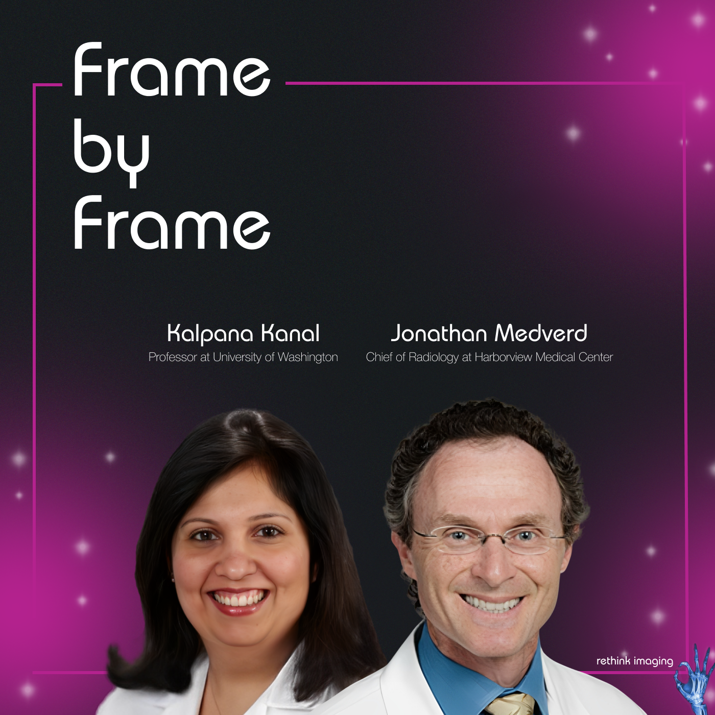
45% Fewer Autopsies: How Postmortem CT Is Changing Death Investigation in the U.S.
Mar 1900:40:13Tap to summarizePostmortem CT is not theoretical. It is operational and growing.In this episode of Rethink Imaging, Dr. Kalpana Kanal and Dr. Jonathan Medverd share the real-world story behind launching a collaborative forensic imaging program between the University of Washington and King County’s Medical Examiner Office. What began as a pilot project six years ago has evolved into a structured clinical pathway that has scanned 184 decedents since July 2022.The results are striking. In roughly 45% of cases, CT findings were sufficient to determine cause of death, eliminating the need for traditional autopsy. In early validation cases, imaging diagnoses including precise identification of a ruptured aorta were confirmed at autopsy, demonstrating strong radiologic-pathologic correlation.Beyond clinical accuracy, the episode dives into the operational realities: aligning two different governance structures, navigating IT firewalls between hospital PACS and county systems, securing funding for scanner installation, building trust between specialties, and operating without a billable CPT code for postmortem imaging. The conversation is candid about volunteerism, resource constraints, and the importance of leadership commitment before launching similar programs.The broader implications are significant. Postmortem CT offers cultural and religious alternatives for families who object to invasive autopsy. It reduces pathologist exposure to infectious diseases and toxic embalming chemicals. It may improve courtroom communication by replacing graphic imagery with diagnostic imaging. And it opens the door to cost efficiencies, particularly when complex autopsies can exceed $2,500 per case.Looking ahead, Dr. Kanal and Dr. Medverd discuss developing a forensic radiology rotation for senior residents and the longer-term vision of a dedicated fellowship combining radiology and forensic pathology expertise. As forensic pathology faces workforce shortages nationwide, hybrid training models may become essential.This episode reframes postmortem CT not as a niche innovation, but as a scalable systems solution with implications for public health, forensic science, radiology training, and healthcare economics.What You’ll Learn:How postmortem CT can replace traditional autopsy in ~45% of casesThe radiologic-pathologic validation process behind virtual autopsyOperational hurdles when partnering between hospital and county systemsHow PACS, DICOM storage, and EMR access were structured across entitiesThe billing and CPT code gap limiting U.S. adoptionCultural and religious considerations in death investigationCost comparisons between CT and complex autopsyWhy forensic radiology fellowships may represent the next evolutionChapters:[00:00] The First Postmortem CT Case That Changed Everything[05:35] Radiology–Medical Examiner Collaboration in Practice[12:07] 184 Cases and a 45% Autopsy Avoidance Rate[18:00] Diagnostic Challenges: Decomposition vs. Trauma[23:29] Building a Forensic Radiology Rotation[29:19] IT, PACS, and Cross-Entity Data Sharing[32:35] Why Billing Codes Are Holding the U.S. Back[34:37] Cost Comparisons and Public Health Impact[36:46] How Counties Can Replicate the ModelRethink Imaging Podcast is handcrafted by our friends over at: fame.so
Transcribe →